 |
Research Article
Platelet transfusion: A study of methods of preparation, storage, quality control, and indications of whole blood-derived platelet concentrates
1 College of Applied Medical Sciences, Al-Dawadmi, Shaqra University, Ministry of Higher Education, Kingdom of Saudi Arabia
2 College of Applied Medical Sciences, Shaqra, Shaqra University, Ministry of Higher Education, Kingdom of Saudi Arabia
3 Department of Laboratory Medicine, Aster Mims Hospital, Kozhikode, Kerala, India
Address correspondence to:
Suhas K Thazha
College of Applied Medical Sciences, Al-Dawadmi, Shaqra University, Ministry of Higher Education,
Kingdom of Saudi Arabia
Message to Corresponding Author
Article ID: 100049Z02ST2019
Access full text article on other devices

Access PDF of article on other devices


How to cite this article
Thazha SK, Scaria B, Mohammed RGA, Rengan SS. Platelet transfusion: A study of methods of preparation, storage, quality control, and indications of whole blood-derived platelet concentrates. Int J Blood Transfus Immunohematol 2019;9:100049Z02ST2019.ABSTRACT
Aims: To study the methods of preparation and storage of whole blood inferred platelet concentrates prepared by the platelet abundant plasma technique, assess the quality control (QC) parameters of whole blood inferred platelet concentrates and study the indications of platelet concentrate transfusion.
Methods: The materials for the present study are a prospective study and were obtained from medico-oncological patients who underwent platelet transfusion therapy with the whole blood-derived platelet concentrates processed by platelet abundant plasma technique in the blood bank of a tertiary care hospital at Kozhikode, Kerala, India during the period of February 2018 to July 2018. The study undertaken in three parts: (1) Study of methods of preparation and storage of whole bloodinduced platelet-rich plasma (PRP) method, (2) Assessment of QC parameters of PRP platelet concentrates, and (3) Study of indications of platelet transfusion therapy in a tertiary care hospital for a period of six months.
Results: In this study, 56 units of whole blood were collected and prepared 56 units of platelet concentrate. One percent of the 56 units prepared was tested of which 75% conformed to the platelet count of not less than 3.5 × 10¹⁰ and 4.5 × 10¹⁰ and 58% of units were found to have a platelet count of more than 4.5 × 10¹⁰. Hundred percent of the volume of platelet units in this study were between 40 and 70 mL and the pH value of all the units were >6.2. The white blood cell (WBC) and red blood cell (RBC) contamination of all the units in this study were less than 1.5 × 10⁹/L and 0.1 × 10¹²/L, respectively. Eighty-six percent of platelet concentrate units in this study were transfused to patients with hematological malignancies and the remaining 19%, 3%, 3% were transfused to nonhematologic malignancies, immune thrombocytopenic purpura, and disseminated infections, respectively.
Conclusion: In conclusion, it was revealed that only minority patients with malignancy required platelet transfusion; nevertheless, platelets were more usually transfused to patients with malignancy than to patients with some other kind of disease.
Keywords: Malignancy, pH, Platelet concentrate units, RBC contamination, WBC contamination, Transfusion
Introduction
Platelet transfusions show an important aspect in the support of hematological, oncological, surgical, and transplant patient. Appropriate utilization of this limited resource commits to most favorable patient care and reduces health care costs.
In the previous years, patients with chronic thrombocytopenia expired of hemorrhage with intensely anticipated number. The increased use of platelet transfusion during the past 15 years has prevented most such deaths. Furthermore, this therapy has permitted increasingly aggressive cytotoxic therapy of malignancy and conversely such therapy has made increasing platelet use necessary.
In any case, in spite of widespread agreement that platelet transfusions contribute hemostasis in thrombocytopenic patients, controversy continues with regard to the optimal dose of platelets to utilize, and the suitable platelet count level at which to transfuse the nonbleeding thrombocytopenic patient [1],[2]. Gmur et al. currently illustrated that a threshold for prophylactic platelet transfusion of platelets could be set at 5000/μL (5 × 10⁹/L) in stable thrombocytopenic patients [3]. According to the protocol, the presence of clinical variables might result in platelet consumption such as fever, bacterial sepsis, splenic pooling, “fresh minor hemorrhages,” and other coagulation disturbances moved the platelet transfusion to bring about 10,000–20,000/μL (10–20 × 109/L) platelets is the usual minimum target level for hemostasis during invasive procedures.
The pertinent benefits of the various techniques for retrieving and storing platelets remain imperceptible. The technique of “platelet rich plasma” and the “buffy coat method” is commonly used to separate platelets from whole blood donations. In addition, apheresisseparated platelets are accepted throughout the world so as to limit donor exposure and to reduce the prevalence of contaminating leukocytes within the preparations. After preparation, platelets are typically kept at 20–24°C [4],[5] within plastic containers whose walls are adequately accessible to oxygen [6],[7] and carbon dioxide. It is best to agitate these preparations regularly. Currently, the storage of platelet concentrate units restricted to five days due to considerations regarding the overgrowth of bacteria which may have inadvertently contaminated the preparation [6],[7]. Activation of platelet in improper maintenance of storage and preparation condition has got direct implication in the quality treatment of the patients.
Quality evaluation of platelet concentrate units ensures the supply of quality blood products for efficient patient care and is important in the laboratory evaluation of platelet transfusion response. Even though majority of platelet transfusions are given to patients whose marrows are suppressed, when the thrombocytopenia is caused by massive blood loss, cardiopulmonary bypass, splenomegaly, immune-mediated thrombocytopenia, and hereditary thrombocytopenia, sometimes platelet transfusion is suggested.
The increasing requirement for platelet transfusions conjoined with the recognition of associated shelf-life problems, bacterial contamination, alloimmunization, and viral diseases transmission has urged to attempts to develop nonviable platelet substitutes [8] and modified autologous erythrocytes [9],[10] capable of enhancing the formation platelet plug.
MATERIALS AND METHODS
The materials for the present study are a prospective study and were obtained in the blood bank of a tertiary care hospital in Kozhikode, Kerala, India during the period of February 2018 to July 2018 from medico-oncological patients who underwent platelet transfusion therapy with the whole blood-derived platelet concentrates processed by PRP technique.
The study is undertaken in three parts:
- Study of methods of preparation and storage of whole blood-derived PRP method.
- Assessment of QC parameters of PRP platelet concentrates.
- Study of indications of platelet transfusion therapy in a tertiary care hospital for a period of six months.
Phlebotomy
Blood collection bags manufactured by the company Terumo Penpol Ltd containing citrate, phosphate, dextrose, and adenine (CPDA) as the anticoagulant were used to prepare whole blood-derived platelet concentrate from a healthy random donor. Before phlebotomy, all donor eligibility criteria were met and also questioned about the history of drug intake likely to affect platelet function, such as aspirin and ticlopidine. In accordance with established standards, phlebotomy was performed. Blood collection volume was 450 ± 10 mL.
The following conditions were seen during phlebotomy:
- Venepuncture was performed by a single prick method.
- Rapid and uninterrupted flow of blood was ensured.
- Blood was constantly mixed with the anticoagulant for 1–2 times/min.
- Ratio of blood and anticoagulant was maintained at a rate of 100:14.
- Phlebotomy was concluded within eight minutes.
- Air entry into the blood bag was prevented by clamping the turbing just near to needle after the needle seats were broken.
Additionally, blood was collected in one ethylenediaminetetraacetic acid (EDTA) and two pilot bottles. The EDTA blood was used to type ABO and Rh, to estimate hemoglobin, and to detect malarial parasites, and the two pilot bottles of blood were used to perform screening tests for human immunodeficiency virus I and II, Hepatitis C virus, Hepatitis B virus, and Treponema pallidum.
Postcollection storage temperature
After the blood collection, the bags were left undisturbed in an air conditioned room for one hour at 23 ± 2°C. In each case, the whole blood units were processed for platelet separation within six hours of phlebotomy.
Preparation of whole blood-derived platelet concentrates by the platelet-rich plasma method
Step 1: Preparation of PRP
Through a light spin (1300 rpm for 13 minutes), whole blood units intended for platelet preparation were placed in diagonally opposite cups of refrigerated centrifuge and temperature set at 20–24°C and then centrifuged these blood bags. The supernatant PRP was transferred to the platelet storage satellite bag and transferred the optimal additive solution (saline, adenine, glucose, mannitol; SAG-M) to the primary bag containing red cells. A dielectric sealer was used to seal the tubing between the primary bag and the satellite bag. After proper labeling, the separate primary bag containing the red cells was then stored at 2–6°C. Then the PRP was processed to yield concentrate of platelets.
Step 2: Preparation of platelet concentrate from PRP
Bags containing PRP were then centrifuged for 15 minutes at 20–24°C at 3000 rpm (heavy spin). The supernatant platelet poor plasma (PPP) was transferred to the second satellite bag, leaving about 40–70 mL of plasma for concentrated platelet resuspension. After labeling as fresh frozen plasma (FFP), the PPP was then stored at 30°C. At room temperature in an air-conditioned room, the bag containing platelet concentrate button was left undisturbed for 1–2 hours. At the end of one hour, the platelets in the resident autologous plasma were evenly resuspended by hand manipulating the platelet storage bags.
Storage of platelet concentrates
In a platelet agitator and incubator employing a horizontal agitation of 70 ± 2 per minute, the platelet concentrates were stored at 20–24°C. The platelet concentrates were stored for 72 hours or five days. It was taken as zero on the first day. Day 1 started the next day at 12 am.
Calibration of refrigerated centrifuge and platelet incubator and agitator
For optimum speeds and spin times for platelet separation, the refrigerated centrifuge was calibrated. The accuracy of the digital thermometer was verified annually in addition to the daily temperature check. The accuracy of speed and time was checked by the hospital’s biomedical engineering department for a period of six months with an accurate tachometer and stop watch. The optimal spin times and speed were also evaluated in conjunction with the evaluation of platelet concentrate QC by function checks.
Regularly calibrated platelet incubators designed to provide a controlled environment for stored platelets and also to accommodate platelet rotators to ensure optimum recovery and function of stored platelets. Temperature recorder charts have been maintained and regularly replaced to ensure the instrument’s temperature range. Also performed were periodic preventive maintenance and cleaning works as well as regular inspection of sensor bottle fluid.
Quality assessment of whole blood-derived platelet concentrates
Prior to release and issue of units, QC parameters of all platelet concentrate units included in this study were evaluated. After hermetically sealing the tube to collect well mixed samples, a representative aliquot of the component was obtained from attached platelet storage bag tubing. After 24 hours of routine storage, the units were selected.
1. Total platelet count of the unit
Platelet count per microliter of platelet concentrate unit was obtained by a five part automated blood cell counter. The amount of platelets present in one unit of platelet concentrate could be easily calculated from platelet count/mL of the unit.
Total number of platelets/unit of platelet concentrate = platelet count/μL of the platelet concentrate × 103 × volume of residual plasma present in the unit.
2. Measurement of pH of platelet concentrate unit
pH of the platelet concentrate unit was measured by using a narrow range pH paper. This method of pH measurement permitted the differentiation of pH values within the range of 5–9.
3. Measurement of volume of platelet concentrate unit
The volume was indirectly calculated by weighing the blood bag using a digital weighing scale.
4. Visual inspection of platelet concentrate unit
It was performed to detect the presence of excessive platelet aggregation and contamination. Units with excessive platelet aggregates were discarded. Pink or red discoloration of the units was a measure of red cell contamination and such units were issued only after performing the cross-matching tests in the unit. Also review the “swirling” or “shimmering” appearance of platelet by holding the bag against a good light source and squeezing it gently.
5. Residual leukocyte and RBC contamination of platelet concentrates
This was estimated by using a five part automated blood cell counter and they were observed by looking at the pulse generated above their lower threshold.
Study of clinical indications for the transfusion of platelet concentrates
The final part of the study examined various conditions for the transfusion of platelet concentrates derived from whole blood.
Statistical analysis
The attained data were possessed and interpreted by SPSS 23 version and the entire data have been expressed as mean ± standard deviation. We applied students t-test to perform statistical comparison. P < 0.05 (two-sided) probability was applied to dismiss null hypothesis.
RESULTS
Quality assessment of platelet concentrates derived from whole blood was prepared by PRP methods and is shown in Table 1.
Seventy-five percent of units were found to have a volume of 60–70 mL. Sixteen percent of the units showed a volume of 50–60 mL and only 9% of the units showed a volume of 40–50 mL. The average volume of the platelet concentrate units was found to be 62 mL (Table 2).
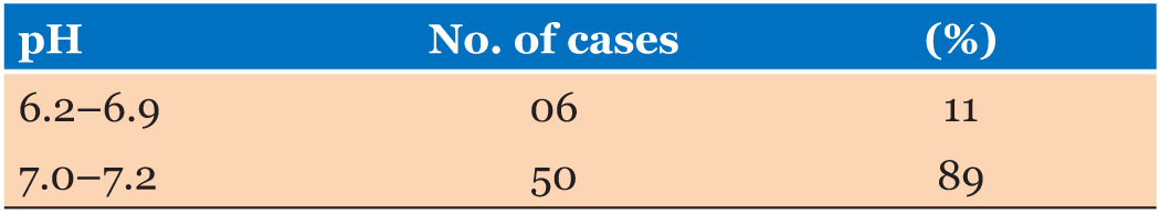
Only 58% of the units were found to have a platelet count of >4.5 × 10¹⁰. Forty-two percent of the units were found to have platelet count in the range of 4.0–4.5 × 10¹⁰. Nine percent of the units had a platelet count of >6 × 10¹⁰. The average platelet count of the units was 4.8 × 10¹⁰ (Table 3). Eighty-nine percent of the platelet concentrate units showed a pH range of 7.0–7.2. Only 11% of the units had shown a pH range of 6.2–6.9. None of the units had shown a pH < 6.2 (Table 4).
On visual examination, none of the units showed the presence of RBC contamination and in the hemogram produced by the automated cell counter. Red blood cell contamination was found to be < 0.05 x 10¹²/L in all the units tested.
White blood cell contamination was significant only in 2% of the units tested, that is >1.5 × 10⁹/L. In 82% of the units, it was found to be < 1.0 × 10⁹/L and in the rest 16% of the tested units, it was present in the range of 1.1–1.5 × 10⁹/L (Table 5).
Visual examination of platelet concentrate units revealed the presence of “swirling appearance” of wellpreserved platelets. It also helped to reveal the absence of excessive platelet aggregates in the platelet concentrate units.
A total of 31 medico-oncological patients constituted the study of clinical indications for platelet transfusion therapy (Figure 1). It was found that management of patients with hematologic malignancies contributed the major clinical indication for platelet concentrate transfusion in medico-oncological patients. Seventy-five percent (23 out of 31 patients) of clinical indications for platelet concentrate transfusion were encountered by patients with hematologic malignancies. Patient management with nonhematologic malignancies contributed only 19% (6 out of 31 patients) of clinical indications of platelet concentrate transfusion. Immune thrombocytopenia (ITP) and disseminated infection were the other two clinical indications for platelet transfusion therapy encountered during this period of study and contributed 3% (2 out of 31 patients) of cases each.
Among the hematologic malignancies, patient management with acute myeloid leukemia (AML) constituted 39% (9 out of 23 cases) of clinical indications for platelet concentrate transfusion. Acute lymphoid leukemia 26% (6 out of 23 cases), non-Hodgkin’s lymphoma 22% (5 out of 23 cases), multiple myeloma 9% (2 out of 23 cases), and chronic myelogenous leukemia 4% (1 out of 23 cases) were the other hematologic malignancies which required platelet transfusion support during this period of study (Figure 1).
Among the nonhematologic malignancies, carcinoma of lung was the major clinical indication for platelet concentrate transfusion and constituted 50% (3 out of 6 patients) of cases. Carcinoma of ovary was the second major reason for platelet concentrate transfusion and constituted 33% (2 out of 6 patients) of cases. Osteosarcoma contributed only 17% (1 out of 6 patients) of cases of platelet concentrate transfusion during this study (Figure 2).


Discussion
The method of “platelet rich plasma” is the most commonly used method of platelet separation. In the present study, the same method was used to prepare platelet concentrates from whole blood.
The entire process of platelet concentrate preparation and storage was checked on a routine basis and followed well standardized techniques as mentioned in the National Legislation on blood and blood products [11]. This study included QC of donor selection, types of container, and anticoagulant preservative solution used for blood collection, the volume of blood collected, the duration of venesection, postcollection storage conditions, the procedures involved in the preparation and storage of components, and lastly the analysis of QC parameters of platelet concentrates before the release and issue of units. The present study satisfied all the abovementioned parameters of platelet concentrate preparation and storage.
The refrigerated blood bank centrifuge and platelet incubator with agitator were used for platelet concentrates preparation and storage. These devices were regularly calibrated with the guidelines mentioned in the National Legislation on blood and blood products. This was done by the biomedical engineering department of the tertiary hospital where I was carrying out the study or by superior in charge of maintenance service contract.
The entire centrifugation process is traumatic to the concentrate platelets. The platelets may aggregate irreversibly and not be functional if they are not allowed to rest. The platelets should therefore be left stationary in the autologous plasma at room temperature (20°C) for about one hour before resuspension. This allows plasma enzyme degradation of platelet adenosine diphosphate (ADP), thus reducing the risk of platelet aggregation [12]. In this study, strict adherence to these criteria was ensured.
Even platelet distribution during platelet concentrate button resuspension was ensured by gentle bag agitation before being placed in the agitator. Failure to agitate the platelets during storage will result in a faster fall in pH and reduced viability through unknown mechanism, independent of pH [13]. This continuous gentle agitation of the platelet concentrates facilitated gas exchange through the walls of the platelet storage bags, plastic containers, and kept the platelets throughout the plasma in an even suspension. The shelf life of platelet concentrate units should be limited to 3–5 days, as a high frequency of septicemia associated with contaminated platelet transfusion has been reported with room temperature storage [12]. In addition, platelets were stored in this study for a period of just three days (First day, i.e., preparation day was taken as zero day. Day 1 began at 12 o’clock on the following day). The present study fulfilled all the criteria of continuous gentle agitation of platelet concentrate units as well as the shelf life mentioned above.
The quality assurance of personnel and premises of the blood bank were also verified in our study. All these steps taken in this study ensured a standardized and wellcalibrated technique of platelet preparation and storage, in the blood bank of tertiary hospital.
Quality control is the process by which product standards are established and met without mistake. The QC parameters ensured the provision of safe transfusion of blood and its components. The World Health Organization (WHO) has defined quality as “the consistent and reliable performance of services or products in conformity with specialized standard” [11]. Since it was not possible to test each individual blood component, at least 1% of the platelet prepared in a month has to be evaluated for QC parameters.
As per the prescribed standards, the volume of units should range from 40 to 70 mL by National Legislation on blood and blood products [11]. The QC criteria were met by 100% of the units included in this study. The volume of original plasma used for platelet resuspension shall be determined during the storage period by maintaining a pH of not less than 6.0. During storage, the platelets metabolize glucose to lactate and hydrogen and are buffered by an adequate supply of bicarbonate in the residual autologous plasma and keep pH above 6.0 for approximately seven days in new plastic containers [14],[15].
pH of platelet concentrate units should be >6.0 at the unit’s storage temperature, according to the prescribed QC parameters. Results from the study showed that all units had a pH value of >6.2, thus meeting the parameters of QC mentioned in the National Legislation on blood and blood products. The decrease in pH due to lactate production from platelet glycolysis is a major limiting factor in storage at 22°C [6]. Also contributing to lesser extent is carbon dioxide released from oxidative platelet phosphorylation [7]. When pH reaches 6.8, the platelet morphology starts to change and it changes dramatically as it reaches 6.0 and loses viability. The rate of pH decline is affected by the number of platelets, the volume of platelets in which they are stored, and the availability of oxygen because oxygen deprivation leads to increased production of lactate [4],[5].
According to National Legislation on blood and blood products, the residual RBC and WBC contamination of the units should be less than 0.1 × 10¹²/L and 1.5 × 10⁹/L, respectively [11]. Results emerged from the study fully satisfied the criteria of QC.
Pursuant to the National Legislation on blood and blood products, the functional viability of platelets should be determined by swirling motion before issuing the units through visual examination [11]. All the units showed a positive “swirling” appearance in the present study and none showed the presence of excessive platelet aggregates.
The time and speed of centrifugation should be shown to produce a count of not less than 3.5 × 10¹⁰ and 4.5 × 10¹⁰, that is platelets per unit from a unit of 350 mL and 450 mL, respectively [11], according to the National Legislation on blood and blood products. One percent of all prepared platelets should be tested, of which 75% should confirm the abovementioned number of platelets [11].
In this study, 56 units of whole blood were collected in blood collection bags of up to 450 mL of triple SAG-M and processed to prepare 56 units of platelet concentrate, as 1% of the 56 units of which 75% conformed to the abovementioned platelet count was tested according to the above criteria. Not only the above criteria have been met, but all the 56 units have also been tested and 58% of units have a platelet count of more than 4.5 × 10¹⁰.
In terms of clinical indications of platelet concentrate transfusion, it has been noted that most units have been transfused to manage patients with hematologic malignancies, according to the previously performed study. In a recent review of practices at the University of Minnesota Hospitals and Clinics, 86% of platelet transfusions were used in patients with hematologic malignancies [16],[17]. Approximately 75% of clinical indications for platelet transfusion therapy contributed to hematologic malignancies in the present study.
According to Bell et al., in patients with solid tumors, the occurrence of thrombocytopenia and hemorrhage was only 10.4% and with platelet counts above 10,000/μL [18] was virtually nonexistent. The results of this study were in agreement with the abovementioned study and only 19% (6 out of 31 cases) cases of platelet transfusion therapy contributed to solid tumors.
Young platelets are more efficient in controlling hemorrhage, thus increasing the need for platelet transfusion if the count drops after chemotherapy compared to similar level during a rise from a nadir. Thus, patients with chronic thrombocytopenia due to reduced platelet production (i.e., myelodysplastic disorders) may require transfusions as opposed to patients with accelerated destruction, but active platelet production (immune thrombocytopenic purpura) [19]. The results of this study were in consistent with this observation. In this tertiary care hospital, only 3% of patients with immune thrombocytopenic purpura required support for platelets transfusion. Disseminated infection during this study period required support for platelet transfusion in 3% of patients.
The thrombocytopenia involves as a hemorrhage risk factor and thus the limitation and dose of prophylactic platelet transfusion may vary in different clinical settings. For example, patients with thrombocytopenia due to AML were reported to have increased bleeding in platelets at ≤10,000/μL as compared to patients with acute lymphoblastic leukemia (ALL) who had a similar risk of hemorrhage in platelets at ≤20,000/μL platelets [20]. The major clinical indication for platelet transfusion during this study period was supported by thrombocytopenia due to AML. Higher platelet counts (40,000–50,000/μL) were required, for example, in patients with high blast counts (>100,000/μL) in the terminal phase of chronic myelogenous leukemia due to the risk of cerebral and pulmonary leukostasis and fatal hemorrhage [20]. During this study period, there was only one case of chronic myeloid leukemia (CML) in the blast crisis phase.
From this study, it was found that only minor cancer patients required platelet transfusion; however, platelets were transfused more frequently to patients with cancer than to patients with any other disease category.
Summary
- All the instruments, equipment, and procedures used in the blood bank and component section for the collection of blood, preparation and storage of platelet concentrate units were as specified by the National Legislation on blood and blood products. These equipment were calibrated regularly on a 3–6 monthly basis and calibration certificates were duly filled.
- The QC parameters mentioned in the National Legislation on blood and blood products were applied to all the platelet concentrate units prepared over a period of four months and it was found that all the units fulfilled all the QC criteria.
- The clinical indications of platelet concentrate transfusion in this tertiary care cancer hospital over a six month period were analyzed and the results were found to be in concordance with the results of most other studies.
Conclusion
In conclusion, it was revealed that only minority patients with malignancy required platelet transfusion; nevertheless, platelets were more usually transfused to patients with malignancy than to patients with some other kind of disease.
REFERENCES
1.
Slichter SJ. Optimizing platelet transfusions in chronically thrombocytopenic patients. Semin Hematol 1998;35(3):269–78.
[Pubmed]

2.
Nahirniak S, Slichter SJ, Tanael S, et al. Guidance on platelet transfusion for patients with hypoproliferative thrombocytopenia. Transfus Med Rev 2015:29(1):3–13. [CrossRef]
[Pubmed]
3.
Gmür J, Burger J, Schanz U, Fehr J, Schaffner A. Safety of stringent prophylactic platelet transfusion policy for patients with acute leukaemia. Lancet 1991;338(8777):1223–6. [CrossRef]
[Pubmed]
4.
Murphy S, Gardner FH. Platelet preservation – effect of storage temperature on maintenance of platelet viability – deleterious effect of refrigerated storage. N Engl Med 1969;280(20):1094–8. [CrossRef]
[Pubmed]
5.
Johnson L, Tan S, Wood B, Davis A, Marks DC. Refrigeration and cryopreservation of platelets differentially affect platelet metabolism and function: A comparison with conventional platelet storage conditions. Transfusion 2016;56(7):1807–18. [CrossRef]
[Pubmed]
6.
Murphy S, Gardner FH. Platelet storage at 22 degrees C: Role of gas transport across plastic containers in maintenance of viability. Blood 1975;46(2):209–18.
[Pubmed]
7.
Murphy S. Platelet storage for transfusion. Semin Hematol 1985;22(3):165–77.
[Pubmed]
8.
McGill M, Fugman DA, Vittorio N, Darrow C. Platelet membrane vesicle reduced microvascular bleeding times in thrombocytopenic rabbits. J Lab Clin Med 1987;109(2):127–33.
[Pubmed]
9.
Collar BS, Springer KT, Beer JH, et al. Thromboerythrocytes. In vitro studies of a potential autologous, semi-artificial alternative to platelet transfusions. J Clin Invest 1992;89(2):546–55. [CrossRef]
[Pubmed]
10.
Behrens MA, Sikorski MJ, Kofinas P. Hemostatic strategies for traumatic and surgical bleeding. J Biomed Mater Res A 2014;102(11):4182–94. [CrossRef]
[Pubmed]
11.
Makroo RN. Compendium of transfusion medicine. Practice of Safe Blood Transfusion 1999;285(98):400.
12.
Mourad N. A Simple method for obtaining platelet concentrates free of aggregates. Transfusion 1968;8(1):48. [CrossRef]
[Pubmed]
13.
Murphy S, Gardner FH. Room temperature storage of platelets. Transfusion 1976;16(1):2–3. [CrossRef]
[Pubmed]
14.
Shahab N, Evans ML. Images in clinical medicine. Platelet satellitism. N Engl J Med 1998;338(9):591. [CrossRef]
[Pubmed]
15.
Kopcinovic LM, Pavic M. Platelet satellitism in a trauma patient. Biochem Med (Zagreb) 2012;22(1):130–4.
[Pubmed]
16.
Gurney AL, Kuang WJ, Xie MH, Malloy BE, Eaton DL, de Sauvage FJ. Genomic structure, chromosomal localization, and conserved alternative splice forms of thrombopoietin. Blood 1995;85(4):981–8.
[Pubmed]
17.
Kuter DJ. The biology of thrombopoietin and thrombopoietin receptor agonists. Int J Hematol 2013;98(1):10–23. [CrossRef]
[Pubmed]
18.
Bell RJ, Leite C, Haas CD, Stephens RL. Incidence of hemorrhagic complications in patients with cancer. JAMA 1978;239(24):2571–4. [CrossRef]
[Pubmed]
19.
Shulman NR, Watkins SP Jr, Itscoitz SB, Students AB. Evidence that the spleen retains the youngest and hemostatically most effective platelets. Trans Assoc Am Physicians 1968;81:302–13.
[Pubmed]
20.
Heyman MR, Schiffer CA. Platelet transfusion to patients receiving chemotherapy. In: Rossi EC, Simon TL, Moss GS, Gould SA, editors. Principles of Transfusion Medicine. 2ed. Baltimore: Williams and Wilkins; 1996. p. 263.
SUPPORTING INFORMATION
Author Contributions
Suhas K Thazha - Conception of the work, Design of the work, Acquisition of data, Analysis of data, Drafting the work, Revising the work critically for important intellectual content, Final approval of the version to be published, Agree to be accountable for all aspects of the work in ensuring that questions related to the accuracy or integrity of any part of the work are appropriately investigated and resolved.
Bibin Scaria - Conception of the work, Design of the work, Acquisition of data, Analysis of data, Drafting the work, Revising the work critically for important intellectual content, Final approval of the version to be published, Agree to be accountable for all aspects of the work in ensuring that questions related to the accuracy or integrity of any part of the work are appropriately investigated and resolved.
Ramieldin GA Mohammed - Conception of the work, Design of the work, Acquisition of data, Analysis of data, Drafting the work, Revising the work critically for important intellectual content, Final approval of the version to be published, Agree to be accountable for all aspects of the work in ensuring that questions related to the accuracy or integrity of any part of the work are appropriately investigated and resolved.
Sameesh S Rengan - Conception of the work, Design of the work, Acquisition of data, Analysis of data, Drafting the work, Revising the work critically for important intellectual content, Final approval of the version to be published, Agree to be accountable for all aspects of the work in ensuring that questions related to the accuracy or integrity of any part of the work are appropriately investigated and resolved.
Guarantor of SubmissionThe corresponding author is the guarantor of submission.
Source of SupportNone
Consent StatementWritten informed consent was obtained from the patient for publication of this article.
Data AvailabilityAll relevant data are within the paper and its Supporting Information files.
Conflict of InterestAuthors declare no conflict of interest.
Copyright© 2019 Suhas K Thazha et al. This article is distributed under the terms of Creative Commons Attribution License which permits unrestricted use, distribution and reproduction in any medium provided the original author(s) and original publisher are properly credited. Please see the copyright policy on the journal website for more information.


